In a groundbreaking study that reads like molecular science fiction, researchers have captured the first high-resolution snapshots of viral capsids undergoing dramatic structural metamorphosis. Using cryo-electron microscopy (cryo-EM), scientists have frozen these shape-shifting pathogens in mid-transformation, revealing secrets about how viruses assemble, infect, and potentially how we might stop them.
The research focuses on what virologists call "viral gymnastics" - the extraordinary structural rearrangements that allow viruses to package their genetic material efficiently before launching infectious attacks on host cells. These observations fundamentally change our understanding of viral architecture dynamics, showing that capsids are far from static molecular containers.
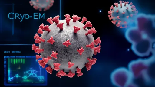
Cryo-EM technology has reached a resolution revolution in recent years, allowing researchers to visualize biological specimens at near-atomic scale. When applied to viral studies, this technique becomes particularly powerful because it captures particles in their native state - frozen in vitreous ice without the distortions caused by chemical fixation or staining. The latest generation of detectors and image processing software can now track structural changes occurring in milliseconds.
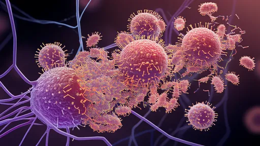
What emerges from these snapshots is a viral world of astonishing flexibility. Capsids - the protein shells that protect viral genomes - were long thought to maintain relatively stable configurations. The new images reveal instead a constant molecular dance where proteins shift, rotate, and reassemble like pieces in an organic jigsaw puzzle. Some viruses appear to completely rebuild their architecture during different life cycle stages.
The most striking observations come from herpesviruses and some bacteriophages, which show radical transformations when transitioning from procapsids to mature virions. These changes aren't gradual tweaks but wholesale reorganizations where hundreds of protein subunits mechanically reposition themselves. It's as if a geodesic dome could spontaneously reconfigure into a completely different polyhedral structure while maintaining structural integrity.
Researchers describe a "molecular breathing" phenomenon where capsid proteins transiently separate and reconnect in new configurations. This flexibility appears crucial for several viral functions: allowing genome packaging, responding to environmental changes, and preparing for host cell entry. The images show viral particles caught in mid-transformation, with some protein subunits already shifted while others remain in their original positions.
One particularly elegant discovery involves the geometric principles governing these transformations. Rather than random motions, the structural changes follow precise rules of quasi-equivalence - a concept in virology where proteins maintain similar bonding patterns while accommodating different curvatures. The cryo-EM snapshots reveal how slight angle changes between subunits enable dramatic overall shape alterations.
These findings have immediate implications for antiviral drug design. Traditional approaches often target static viral structures, but the new understanding suggests we might develop compounds that interfere with structural transitions instead. Imagine a drug that freezes a virus in its pre-assembly state or prevents it from achieving the configuration needed for cell entry. Several research groups are already exploring this strategy.
The technological achievements behind this research deserve special mention. Capturing viral metamorphosis required not just advanced cryo-EM equipment but innovative sample preparation techniques. Scientists developed rapid-freezing methods that can trap transient structural states lasting mere milliseconds. Combined with computational sorting algorithms that can classify subtle conformational differences among thousands of particles, these advances created molecular movies from what were once static snapshots.
Beyond medical applications, the research offers insights into fundamental questions about self-assembling systems. Viruses demonstrate how complex structures can build themselves through simple interaction rules between components. Materials scientists are particularly interested in how viral proteins achieve such precise large-scale rearrangements without external energy input - a potential blueprint for designing adaptive nanomaterials.
As with any major advance, the discoveries raise new questions. How do viral genomes influence capsid transformations? What signals trigger specific structural changes? Can we predict all possible configurations a given virus might adopt? The research teams are now developing time-resolved cryo-EM techniques to address these mysteries, along with advanced computational simulations that can model the dynamic energy landscapes of viral proteins.
The study also provides a cautionary note about virus variability. The observed structural flexibility may help explain why some viruses evade immune responses so effectively - their surface proteins can shift to avoid antibody recognition while maintaining core functions. This adaptability presents challenges for vaccine development but also suggests new targets for broad-spectrum antivirals.
Looking ahead, researchers anticipate cryo-EM will reveal even more surprising viral behaviors. Early work suggests some viruses may have multiple stable configurations rather than a single "correct" structure. Others appear to sample different shapes transiently before settling into their infectious form. These observations blur the line between structural biology and dynamic systems theory, inviting interdisciplinary approaches to virology.
The viral shape-shifting captured in these studies resembles a microscopic version of metamorphosis seen in insects or amphibians, but occurring through molecular mechanics rather than cellular differentiation. It's a reminder that evolution has produced remarkable nanoscale machines in viruses - entities that exist on the boundary between chemistry and biology, capable of radical transformations that would challenge human engineers.
As cryo-EM technology continues advancing, scientists expect to visualize even faster and subtler viral dynamics. The next frontier involves combining these structural insights with functional assays to understand how each conformational change relates to infectivity. This integrated approach may finally reveal the complete choreography of viral infection - from first host contact to genome release.
For now, the research stands as a testament to how cutting-edge technology can transform our understanding of fundamental biological processes. What once seemed like stable viral structures are now understood as dynamic ensembles, constantly shifting between configurations like molecular origami. This paradigm shift will undoubtedly influence virology for decades to come, from basic research to clinical applications.
By /Aug 14, 2025

By /Aug 14, 2025
By /Aug 14, 2025
By /Aug 14, 2025
By /Aug 14, 2025

By /Aug 14, 2025

By /Aug 14, 2025

By /Aug 14, 2025

By /Aug 14, 2025
By /Aug 14, 2025
By /Aug 14, 2025

By /Aug 14, 2025
By /Aug 14, 2025

By /Aug 14, 2025
By /Aug 14, 2025
By /Aug 14, 2025

By /Aug 14, 2025

By /Aug 14, 2025

By /Aug 14, 2025
By /Aug 14, 2025